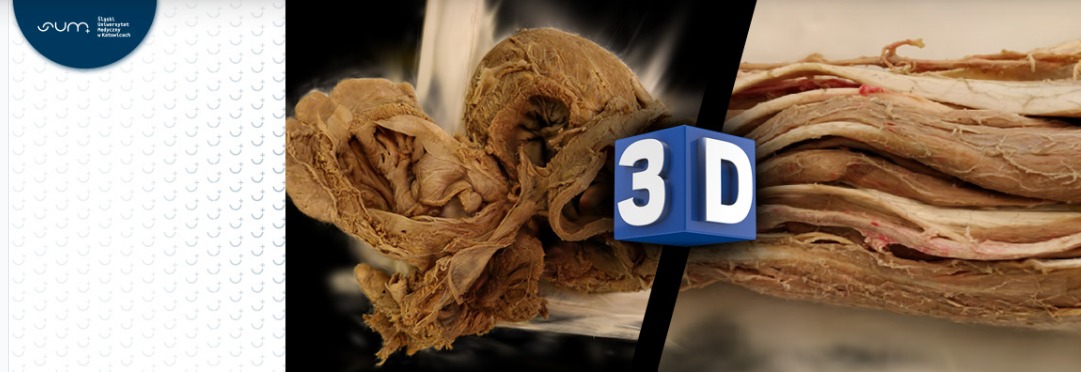
The Silesian Medical University has made it possible for students to practise anatomy using a set of high-quality and high-fidelity 3D models prepared from photographs of the actual specimens they are studying in class. Artificial intelligence helped to achieve the effect. This educational revolution will definitely make it easier to pass this challenging exam.
A set of 3D images of the preparations, including upper limbs, lower limbs, thorax and a number of preparations in line with the syllabus were prepared by Przemysław Jędrusik’s team, head of the SUM Centre for Remote Learning and Educational Effects Analysis, specifically for teaching anatomy. – The students also tell us which preparation is the most difficult for them and this is the one we prepare first. They will be able to see it in detail and learn without leaving home, because we give them the opportunity to use our preparations on their own computers. Not just in the library,’ explains Jędrusik and adds: – 'Designed and produced such an advanced viewer of anatomical preparations at this level of fidelity is the only one of its kind in Poland. We have prepared a didactic tool redefining the teaching process for students through independent access to materials in a quality that has not been available before. Of course, our collection will continuously grow.
Anatomy is considered one of the most difficult subjects. During colloquia and examinations, students have to demonstrate flawless theoretical and, above all, practical knowledge of the human body, which involves recognising its specific structures marked with a so-called „pin”. (Particularly difficult are those related to the structure of the Central Nervous System.) During classes, students use preparations after extracting them from the formalin that fixes them.
– Our project is the development and implementation of a supporting educational tool for students, transferring the acquisition of anatomical knowledge to a completely new dimension with the use of artificial intelligence and optimisation algorithms,” says Przemysław Jędrusik, who evaluates: – 'The development of modern technologies enables the extensive use of high-performance algorithms also in areas related to medicine. Based on an innovative approach of mapping the visualisation of scenes recorded using a series of photographs, we have developed a teaching tool in the form of a viewer for high-resolution, 3D models of anatomical preparations.
The method used is based on 3D Gaussian Splatting (3D Gaussian Splatting) of image transformation, where the 3D space is defined as a Gaussian set whose element parameters are calculated using machine learning.
– The whole process enables real-time transformation of photorealistic scenes from small samples of images.
„3D Gaussian Splatting presents each scene as a point cloud estimated from a set of images, converted into a set of millions of particles (Gaussians), where each element of the set has a specific position, orientation, scale, colour and transparency, resulting in an image that so faithfully reflects real preparations,” explains Jedrusik.
In general, we cannot tell the difference between the preparation from the computer screen, which was created with the support of artificial intelligence, and the one in the dish with formalin.
The high quality of the results obtained in 3D space is made possible through the use of machine learning, using an optimisation algorithm. The algorithm ultimately allows the elements of the Gaussian set to better match fine-grained details, while removing redundant, unnecessary elements of the set. We thus have at our disposal a product that almost perfectly reflects reality. – The prepared repository allows each student to rework and consolidate the knowledge acquired in traditional classes in the dissection rooms at their own pace and at any time, based on the materials used in class. The faithful reproduction of real specimens in the form of 3D models, which can be freely zoomed, rotated and moved, significantly reduces learning time, while maximising the knowledge acquired in anatomy,” says Przemysław Jędrusik.
It is worth adding that the usefulness of traditional slides is short. They can usually be used for up to a year when used in classes for groups of students twice a week. At the Silesian Medical University, a programme for conscious cadaver donation was established in 2003. Today, the SUM Department of Normal Anatomy has more than 2,000 notarised deeds from people whose will is to donate a body to SUM for scientific purposes after death.
Undoubtedly, the new educational tool of 3D images will definitely facilitate learning.

